Bullous Pemphigoid

A-Z OF SKIN

Bullous Pemphigoid

BACK TO A-Z SEARCH

Last updated: January 2024

What is bullous pemphigoid?

Bullous pemphigoid is a skin condition where the immune system mistakenly attacks the skin and causes blisters to form. The term “bullous” refers to these blisters and “pemphigoid” comes from the Greek word pemphix and means bubbles.

Bullous pemphigoid is the most common type of autoimmune blistering disease.

Who gets bullous pemphigoid?

Bullous pemphigoid can occur at any age, but it is more common in individuals over 70 years of age.

In rare cases, bullous pemphigoid has been reported in infants and children.

Individuals with bullous pemphigoid are about five times more likely to have a neurological disease such as dementia, Parkinson’s disease and cerebrovascular disease. 1 Other things that are linked to bullous pemphigoid include malignancy, psoriasis and diabetes mellitus.

What causes bullous pemphigoid?

There are two main layers of the skin, the top layer is called the epidermis and the deeper layer is the dermis. The area between these layers is known as the basement membrane zone. In bullous pemphigoid, the body’s immune system produces proteins (autoantibodies) which attack and damage specific proteins in the basement membrane zone (known as BP180 and BP230). These proteins are critical in attaching the top layer of skin cells (the epidermis) to the underlying layer (the dermis). 

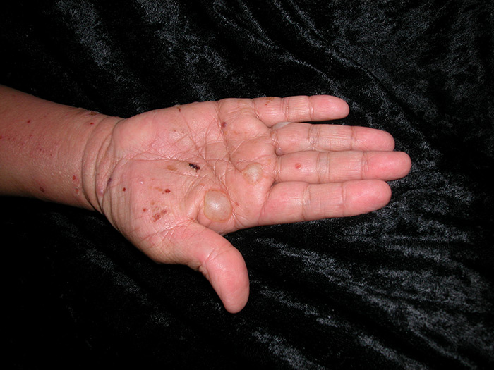
In bullous pemphigoid, the basement membrane proteins are damaged, which causes the layers of the skin to separate, leading to blister formation.

What causes the immune attack on the skin is unknown. Bullous pemphigoid sometimes occurs after an infection or, in a few cases, it occurs after taking new medications. Medications with the strongest associations include gliptins, checkpoint inhibitors, loop diuretics, and penicillins.

What does bullous pemphigoid look like?

Bullous pemphigoid can initially look like dermatitis or urticaria (hives) with red itchy bumps and patches. Unlike hives, these patches do not move around or disappear. The condition may stay like this for months until blisters eventually start to form.

The blisters are typically large, tense, fluid filled and very itchy.  They occur most frequently on the skin of the body folds and trunk. Usually, the condition does not affect inside the mouth.

BullousPemphigoid

Figure 1. Image reproduced with permission of Prof. Dedee Murrell

BullousPemphigoid

Figure 2. Image reproduced with permission of Prof. Dedee Murrell

BullousPemphigoid

Figure 3. Image reproduced with permission of Prof. Dedee Murrell

How is bullous pemphigoid diagnosed?

Because there usually are no blisters initially, diagnosing bullous pemphigoid may be delayed.

The following tests are usually required:

  1. Skin biopsies: This involves taking a small piece of skin, usually from the edge of a blister for routine pathology. A second biopsy is also taken from the skin close to a blister and is checked for autoantibodies with a special technique called direct immunofluorescence.
  2. Blood tests: These test for autoantibodies in the blood.

The Bullous Pemphigoid Area Index (BPDAI) is used to determine the severity of an individual’s bullous pemphigoid and best treatment. It also helps assess the severity and activity of the disease and can track the disease over time. Autoimmune Bullous Disease Quality of Life (ABQOL) is another tool used to measure how much the disease is impacting an individual’s daily life.

How is bullous pemphigoid treated?

Treatment options will vary depending on the individual and their needs.

Bullous pemphigoid treatment aims to reduce blister formation, relieve pruritus (itchiness), promote wound healing and minimise the side effects of treatment. Treatment options may include:

Avoiding triggers

When a drug cause is suspected, the suspected drug culprit should be discontinued where feasible.

Mild Disease

  • Potent topical corticosteroid
  • Tetracyclines (i.e., Doxycycline), with nicotinamide

More aggressive disease

Oral medications, such as:

  • First-line treatment e.g., oral prednisolone
  • Second-line treatments e.g., topical immunomodulators (e.g. tacrolimus), dapsone, azathioprine, mycophenolate, methotrexate

Refractory disease (resistant to treatment)

  • Intravenous immunoglobulin (IVIG)
  • Rituximab

What is the likely outcome of bullous pemphigoid?

Bullous pemphigoid is usually a chronic disease that may have recurrent flares over many years. For some individuals, it can be resolved after a few years. 

  1. Lai YC, Yew YW, Lambert WC. Bullous pemphigoid and its association with neurological diseases: a systematic review and meta‐analysis. Journal of the European Academy of Dermatology and Venereology. 2016 Dec;30(12):2007-15.

Dr Cathy Zhao, Dr Ben Koszegi and Professor Dedee Murrell

January 2024

Dr Cathy Zhao, Dr Belinda Welsh, Dr Ben Koszegi and Professor Dedee Murrell

February 2020

Disclaimer

2019 © Australasian College of Dermatologists.

You may use for personal use only. Please refer to our disclaimer.