What is mastocytosis?
Mastocytosis is a condition where mast cells accumulate in the skin or sometimes in internal organs.
Mast cells are normal cells in the body, usually found in the skin and other tissues. Mast cells are part of our immune system and have a role in the body’s ability to fight certain infections (mainly parasites) and the body’s coordination of healing responses to an injury and reaction to an allergy.
Mast cells contain many different chemicals, such as histamine, that increase inflammation. In the skin this results in localised itching, swelling, redness and sometimes blistering (similar to what happens normally in insect bites).
What causes mastocytosis?
Most forms of mastocytosis are caused by a change in a single area of a gene (c-kit) that causes overproduction of mast cells. In most cases there is no family history of mastocytosis and it is rarely, if ever, passed on to the next generation.
What does mastocytosis look like?
Mastocytosis most commonly affects only the skin where it is referred to as “cutaneous mastocytosis”. The condition can also involve internal organs where it is referred to as “systemic mastocytosis”. There are some cases of systemic mastocytosis which do not have skin involvement.
Cutaneous mastocytosis
Cutaneous mastocytosis causes itching, swelling and blistering of the affected skin, particularly when it is rubbed or scratched.
There are two main types of skin mastocytosis:
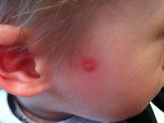
- Solitary mastocytoma
This is a single reddish or yellowish-brown thickened patch of skin. It usually occurs in infants and resolves on its own over the first decade of life.

- Maculopapular cutaneous mastocytosis (previously known as urticaria pigmentosa)
This is a rash with light brown, itchy, raised patches on any part of the body. This form can affect either children or adults. When children are affected, it tends to occur while they are quite young and usually resolves around puberty. When it starts in older children or in adults, it tends to be more persistent and can be associated in some cases with internal involvement and/or systemic symptoms. Internal involvement is very rare in young children.
There are less common forms of skin mastocytosis:
- Diffuse cutaneous mastocytosis
This usually occurs in the first year of life. It presents with widespread red and itchy skin that may blister.
- Telangiectatic cutaneous mastocytosis(previously called telangiectasia macularis eruptiva perstans)
This is an adult type of mastocytosis. It is very persistent and may sometimes lead to systemic involvement.
Systemic mastocytosis
Systemic mastocytosis refers to an increase in mast cells in internal organs. The majority of cases will have non-skin (systemic) symptoms. There may or may not be skin involvement as well. A wide range of symptoms can occur depending on where the mast cells are. The vast majority of people with systemic mastocytosis have a form called indolent systemic mastocytosis.
The severity of symptoms depends on the number of mast cells in the tissues. A high load of mast cells tends to lead to more severe symptoms.
Non skin symptoms can include flushing, headache, abdominal pain, diarrhoea, nausea, vomiting, racing of the heart, bone aches, anxiety, shortness of breath, wheezing and fainting.
How is mastocytosis diagnosed?
Cutaneous mastocytosis is usually diagnosed by its appearance and the fact that rubbing the affected area causes redness or even blistering (known as Darier’s sign). A skin biopsy may be helpful to confirm the diagnosis.
A blood test showing levels of tryptase 5 to10 times the upper limit of normal may suggest systemic involvement.
Some people may need extra tests to check for systemic involvement such as blood tests, scans and, in some cases, a bone marrow biopsy.
How is mastocytosis treated?
Most localised forms of cutaneous mastocytosis (solitary mastocytoma) without symptoms require no treatment.
The vast majority of people with cutaneous mastocytosis, including those with systemic mastocytosis, have a normal life expectancy and treatment is directed at symptom control.
General measures
If there is systemic involvement, certain triggers may cause symptoms to occur. Not all triggers occur in all people with systemic symptoms. Triggers should be avoided.
Possible triggers for mastocytosis symptoms include:
- Alcohol
- Physical stimuli (such as heat, cold, friction or pressure on skin, sunlight, fatigue, exercise)
- Bites and stings (such as insect bites, jellyfish stings, snake bites)
- Infection with fever (bacterial, fungal or viral)
- Drugs including:
- General anaesthetics (such as neuromuscular blocking agents, opiates)
- Pain relief medications (such as aspirin, NSAIDs, morphine, codeine)
- Cough syrup
- Certain local anaesthetics (such as procaine)
- Certain antibiotics (such as vancomycin, polymyxin B, amphotericin)
- Certain blood pressure medications (such as alpha and beta blockers)
- Dextran, dextromethorphan, quinine
- Some radiographic dyes, particularly those containing iodine
Specific treatments
The treatment needed depends on an individual’s symptoms and test results. It may include the following:
- Antihistamines often at high doses (up to 4 times the normal dosage)
- H2 histamine antagonists (such as ranitidine), proton pump inhibitors (such as omeprazole, lansoprazole, pantoprazole) for gastrointestinal symptoms
- Intramuscular adrenaline/epinephrine (EpiPen®) should be used for anaphylactic reactions
- Oral steroids may be used in some cases of systemic mastocytosis
- Light therapy (phototherapy) has been used for localised cutaneous mastocytosis in some cases
Severe cases of systemic mastocytosis or aggressive forms of mastocytosis may be treated with systemic medications, including types of chemotherapy or immune therapy.
This information has been written by Dr Narelle Bleasel
Updated 22 June 2015